Minimally Invasive Spine Surgery (MIS)
When compared to the more traditional open surgery done with really long incisions, minimally invasive spine surgery approaches can be faster, safer, and require less recovery time. Many times, the surgery can be done using incisions several millimeters or less.
Because of the reduced damage to the muscles and soft tissues during surgery, surgeries performed with MIS techniques can:
- Have better cosmetic results because the incisions are smaller
- Less scar tissue
- Less blood loss
- Less time under anesthesia
- Reduced risk of muscle damage (since no cutting of the muscle is done and instead the muscle is moved to the side).
- Faster recovery
- Less rehabilitation is required
Frequently Asked Questions
What conditions are generally treated in a minimally invasive manner?
There are a number of various conditions that can be treated via a MIS approach including:
- Degenerative disease disease
- Herniated discs
- Sacroiliac (SI joint) Dysfunction
- Vertebral compression fractures
- Lumbar spinal stenosis
- Lumbar spondylithesis
How does Minimally Invasive Surgery work?
Traditionally, surgeons used very long incisions to perform spine surgery requiring patients to stay in the hospital for days to weeks after surgery. Patients who underwent these surgeries were at risk for postoperative complications including infection, prolonged hospital stays, increased bleeding, increased risk of blood clots, and more pain following surgery. With advancing technology and techniques, we are able to perform surgery in the most minimally invasive fashion using very small incisions and therefore less muscle and soft tissue damage. Therefore, patients generally leave the hospital sooner, have less blood loss, and recover quicker.
What are some of the common procedures treated with minimally invasive techniques?
Disc herniations: If you are a candidate, Dr. Webb can perform your surgery through a small tube and very small incision. This procedure, generally done through a 22mm tube, is also called a MITR (minimally invasive tubular retraction). This surgery involves progressive dilation of your soft tissues as opposed to cutting them. By using the tubes to move the muscles out of the way, Dr. Webb is able to work through a small incision without having to expose an entire part of your spine. Once the surgery is complete, the tubular retractor is removed, allowing the dilated tissues and muscles to fall back into place. By using the tubes to perform your surgery in this minimally invasive fashion, your recovery is quicker, you may need less pain meds after surgery, your infection risk is less, and you will be able to get back to doing some of the things that you enjoy doing quicker.
Lumbar Spinal Stenosis: If you are a candidate, Dr. Webb may sometimes perform your decompression surgery through a small tube via a very small incision. This procedure, generally done through a 22mm tube, is also called a MITR (minimally invasive tubular retraction). This surgery involves progressive dilation of your soft tissues as opposed to cutting them. By using the tubes to move the muscles out of the way, Dr. Webb is able to work through a small incision without having to expose the area widely. Once the surgery is complete, the tubular retractor is removed, allowing the dilated tissues and muscles to fall back into place. By using the tubes to perform your surgery in this minimally invasive fashion, your recovery is quicker, you may require less pain meds after surgery, your infection risk is less, and you will be able to get back to doing some of the things that you enjoy doing quicker.
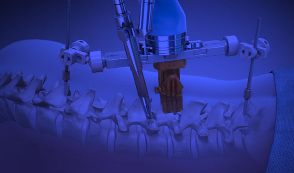
Posterior Lumbar fusions: If you are a candidate, Dr. Webb may be able to perform your lumbar fusion through very small incisions on your lower back. Through these small incisions, Dr Webb will perform percutaneous placement of percutaneous screws and rods that will help fuse and stabilize your spine. Percutaneous (meaning through the skin) placement of the screws and rods is done with x-ray guidance to ensure accuracy and precision of insertion. Traditionally, the incisions for lumbar fusions required extensive dissection of muscle and soft tissue in order to complete the surgery. Using minimally invasive techniques, patients generally recover quicker, have shorter hospital stays, less blood loss, and ultimately return to their activities sooner.
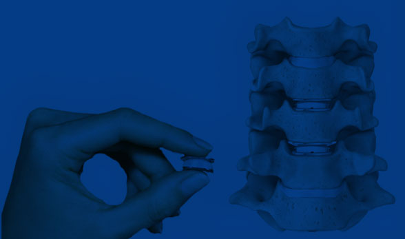
Lateral Lumbar fusions: If you are a candidate for this procedure, Dr. Webb may be able to perform your lateral lumbar fusion through a very small incision on the side of your abdomen. After the diseased disc is removed, Dr. Webb will then insert a metal or plastic cage filled with bone graft into this area. This procedure may also involve turning you prone (after Dr. Webb is done with the lateral part of your surgery) and then placement of percutaneous screws and rods that will help fuse and stabilize your spine. Percutaneous (meaning through the skin) placement of the screws and rods is done with x-ray guidance to ensure accuracy and precision of insertion. Traditionally, the incisions for lumbar fusions required extensive dissection of muscle and soft tissue in order to complete the surgery. Using minimally invasive techniques, patients generally recover quicker, have shorter hospital stays, less blood loss, and ultimately return to their activities sooner.
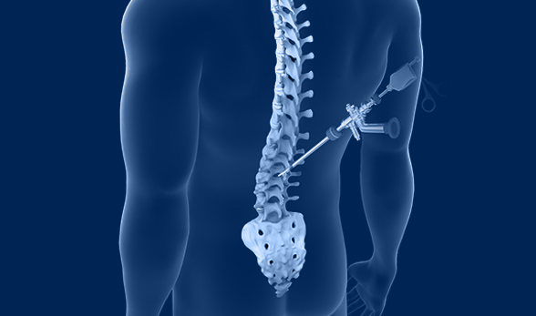
Sacroiliac (SI) Joint Fusion: If the SI joint is the cause of your low back pain, you may be a candidate for a minimally invasive SI joint fusion. This is usually an outpatient procedure that involves carefully placing metal screws across the SI joint, so the bones fuse and grow together. Fusing the joint helps to alleviate the pain associated with excess movement. This procedure is generally done through a very small incision and patients usually go home the same day of surgery.